
Cross Section Analysis
Sample Preparation
A group of sample fragments were received by mail, and two larger fragments were mounted in Ward’s Bioplast mounting medium analysis (a polyester/methacrylate blend) for cross sectional analysis, polymerized with the addition of methylethylketone peroxide, and cured in an illuminated oven at 60 deg. C for three hours under a 6OW tungsten halogen bulb. The embedded samples were initially sectioned on a belt type of sander, and prepared (dry) with the use of progressively finer bonded abrasive clothes, to a grit of 12,000, from Micro-Mesh Inc. The samples were coverslipped using mineral spirits prior to examination.
Images of the cross-sectioned samples were recorded on Kodacolor 200 print film using a Nikon Labophot microscope equipped for epi-illumination in the UV and in normal light. The magnification for each sample view is 120x unless otherwise noted. The illumination conditions were, for the UV light shots; a 360-430 nm excitation, 430 nm suppression filter, from an HBO 100 W mercury source. For the normal light image, a tungstenhalogen lamp (15W, Osram) was used -without any additional color correction, but with cross polarization.
Sectional views of the samples were photographed in any or all of the following conditions; normal light (‘N’), UV only (‘UV’), UV light stained with with 4% Triphenyl Tetrazolium Chloride in methanol (TTC’, a general carbohydrate stain), .2% Rodamine B in ethanol (‘RHOB’, a general lipid stain); and .2% Flurescamine in acetone (FLUR’, a general protein stain).
Results from Sample Examination
Sample from Lunette of Food and Fiber Building
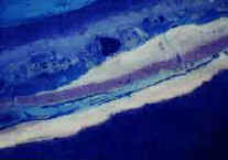
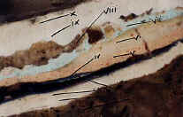 The sample appears to begin with a masonry substrate (i); the surface to the masonry appears to have been primed with two applications of a white emulsion type primer (ii a, b) (bright autofluorescence, +TTC, +FLUR, +RHOB). Applied over this priming is a single layer of blue paint (iii) (dark- autofluorescence; +RHOB), that appears to carry a natural resin varnish (iv), darkened and discolored with exposure, as the original presentation surface.
 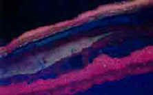 The surface to (iv) is soiled. Next in sequence appears a pinkish oil overpaint layer (v) (dark autofluorescence; +RHOB; soiled surface). Then next in sequence an offwhite priming (vi) (bright autofluorescence, surface +FLUR), and a blue top coat (vii) (dark autofluorescence, +RHOB). Then what appears to be an aggregate containing “texture” paint (viii) (dark autofluorescence; non-staining) is next; followed by a white synthetic paint (ix)(dark autofluorescence, +RHOB), and what appears to be the current presentation coating, a tan synthetic (x)(dark autofluorescence, +RHOB).
Layer (iii) : Closest Match; See Benjamin Moore 734
L 50. 34 a -31-17 b -7.83
 NOTE: The layers (viii-x) are clearly synthetic, post world war paints. The initial presentation sequence (ii-iv) could certainly fall between the wars as an oil-over-emulsion priming type decorative sequence. As I recall, this sequence compares very similarly to that noted in the earlier fair park samples.—-RCW
Click Here to view the main report for this project in Adobe PDF format
(if you need the free Adobe Acrobat reader, click this link http://www.adobe.com/products/acrobat/readstep2.html )
Click here to view the cross section photographs for the main report.